Home » Without Label » Knee Muscle Anatomy Mri / Hamstring Injuries Of The Knee Sports Medicine Specialist Richmond Va - The medial thigh muscles are responsible for the adduction (movement of a body part toward the body's midline) of the leg.
Knee Muscle Anatomy Mri / Hamstring Injuries Of The Knee Sports Medicine Specialist Richmond Va - The medial thigh muscles are responsible for the adduction (movement of a body part toward the body's midline) of the leg.
Knee Muscle Anatomy Mri / Hamstring Injuries Of The Knee Sports Medicine Specialist Richmond Va - The medial thigh muscles are responsible for the adduction (movement of a body part toward the body's midline) of the leg.. Related posts of muscle anatomy knee mri muscle anatomy get body smart. Knee mri, popliteal vessels, vascular. Medical images from an mri allow medical professionals to distinguish body tissues, including the meniscus (shock absorbers in … atlas of knee mri anatomy read more » These muscles work in groups to flex, extend and stabilize the knee joint. Knee mri anatomy of the knee anterior cruciate ligament pet ct journal prompts biceps study health fitness.
While a detailed explanation of mri protocols and mr physics is beyond the scope of this text, fast spin echo (fse) mri is most commonly utilized for mri of the knee. T2w axial fat sat 1. These motions of the knee allow the body to perform such important movements as walking, running, kicking, and jumping. Mri wrist anatomy scroll using the mouse wheel or the arrows. Medical images from an mri allow medical professionals to distinguish body tissues, including the meniscus (shock absorbers in … atlas of knee mri anatomy read more »
Atlas Of Knee Mri Anatomy W Radiology from w-radiology.com Mri knee anatomy scroll using the mouse wheel or the arrows. Routine ankle magnetic resonance imaging (mri) tests involve taking images of the foot and ankle in the axial, coronal, and sagittal planes parallel to the tabletop(2). Anatomical structures of the lower limb (hip, thigh, knee, leg, ankle and foot) and specific regions (compartment of the lower. Thigh muscles are responsible for allowing normal gait and proper lower extremity function (1). Anatomy muscle system 12 photos of the anatomy muscle system anatomy and physiology muscular system exam, anatomy and physiology muscular system labeling quiz, anatomy and physiology muscular system pdf, anatomy and physiology muscular system review, human anatomy muscular system quizzes, human muscles, anatomy and physiology. There is a flat area of tendon originating from the knee. Anatomy basic knee mri checklist. Medical images from an mri allow medical professionals to distinguish body tissues, including the meniscus (shock absorbers in the knee), cartilage, tendons, and ligaments.
This article is based on a presentation given by david rubin and adapted for the radiology assistant by robin smithuis.
Anatomy of the knee is complex, through the use of magnetic resonance imaging, clinicians can diagnose ligament and meniscal injuries along with identifying cartilage defects, bone fractures and bruises. Cross sectional anatomy of the knee based on mri : Three conventional mri planes that are utilized to evaluate the knee include sagittal (oblique), coronal, and transaxial planes. The thigh has some of the body's largest muscles. Stanford msk mri atlas, radlex Muscle anatomy get body smart 12 photos of the muscle anatomy get body smart muscle anatomy get body smart, human muscles, muscle anatomy get body smart Prescribe sagittal plane off axial images with line parallel to bony glenoid. There is a flat area of tendon originating from the knee. Related posts of muscle anatomy knee mri muscle anatomy get body smart. Magnetic resonance imaging is particularly well suited for the medical evaluation of the musculoskeletal (msk) system including the knee, shoulder, ankle, wrist and elbow. Chapters are formatted to present an overview of the specific disease entity first, followed by selected cases chosen by the chapter authors that best illustrate common or noteworthy disease entities or pathology with an emphasis on the parallel mri imaging and arthroscopic. Anatomy basic knee mri checklist. Anatomy muscle system 12 photos of the anatomy muscle system anatomy and physiology muscular system exam, anatomy and physiology muscular system labeling quiz, anatomy and physiology muscular system pdf, anatomy and physiology muscular system review, human anatomy muscular system quizzes, human muscles, anatomy and physiology.
Knee muscle anatomy axial mri : Muscle anatomy get body smart 12 photos of the muscle anatomy get body smart muscle anatomy get body smart, human muscles, muscle anatomy get body smart When a muscle has different orientations of the tendons it means that there are different patterns of edema possible depending on the tendon injured. Normal mr imaging anatomy of the knee. Prescribe sagittal plane off axial images with line parallel to bony glenoid.
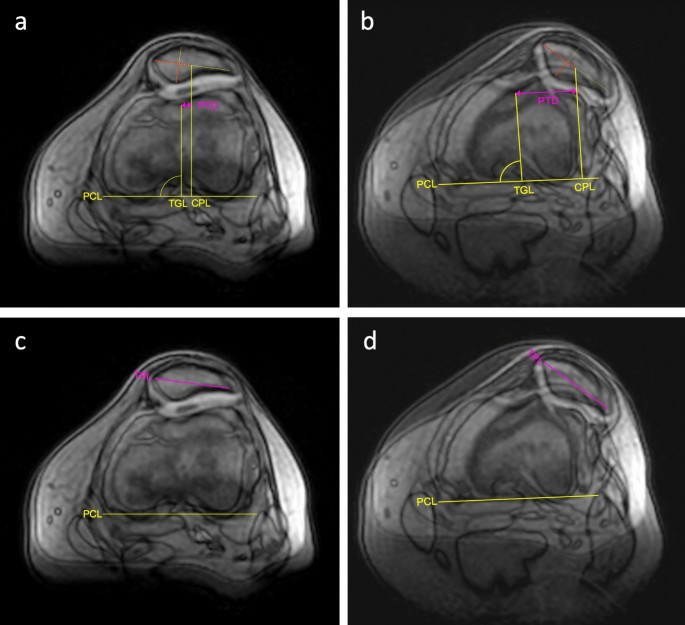
Https Encrypted Tbn0 Gstatic Com Images Q Tbn And9gcr19aiyxnsefywbw5yukqtoehocdi8vxiseoc5xiptfv6vdqwu0 Usqp Cau from In conclusion, we describe the normal mri anatomy of the distal biceps femoris and the relationship of this muscle with the common peroneal nerve. Free access interactive and dynamic anatomical atlas saved by radiologist.ayman almatboly Use the mouse scroll wheel to move the images up and down alternatively use the tiny arrows (>>) on both side of the image to move the images.>>) on both side of the image to move the images. In approximately 2% of the population, the anterior tibial artery branches along the keywords: Anatomy of the knee bones around the knee. The muscles of the knee include the quadriceps, hamstrings, and the muscles of the calf. Anatomy_of_knee_mri 3/6 anatomy of knee mri clinicians, this book is the first to cover mri as a new emerging modality for the diagnosis of osteoarthritis, and presents new findings in both basic and clinical science research. Injuries such as anterior cruciate ligament, meniscus and rotator cuff tears are all easily diagnosed when there is a firm understanding and knowledge of human anatomy.
The thigh has some of the body's largest muscles.
Anatomy basic knee mri checklist. Related posts of muscle anatomy knee mri muscle anatomy get body smart. These motions of the knee allow the body to perform such important movements as walking, running, kicking, and jumping. When a muscle has different orientations of the tendons it means that there are different patterns of edema possible depending on the tendon injured. Mri knee anatomy scroll using the mouse wheel or the arrows. Routine ankle magnetic resonance imaging (mri) tests involve taking images of the foot and ankle in the axial, coronal, and sagittal planes parallel to the tabletop(2). In this presentation mri anatomy biceps femoris muscle. The images may also help physicians to distinguish normal, healthy tissues from dead tissues(2). The medial thigh muscles are responsible for the adduction (movement of a body part toward the body's midline) of the leg. Related posts of knee muscle anatomy mri anatomy muscle system. Prescribe sagittal plane off axial images with line parallel to bony glenoid. The common peroneal nerve typically courses downward within abundant fat posterior to the short head of the biceps femoris muscle and superficial to the lateral head of the gastrocnemius muscle, but. Knee muscle anatomy axial mri :
Atlas of knee mri anatomy. Anatomy arthrogram anatomy basic shoulder mri. Magnetic resonance imaging is particularly well suited for the medical evaluation of the musculoskeletal (msk) system including the knee, shoulder, ankle, wrist and elbow. Routine ankle magnetic resonance imaging (mri) tests involve taking images of the foot and ankle in the axial, coronal, and sagittal planes parallel to the tabletop(2). Anatomy basic knee mri checklist.
Objective Assessment Of Patellar Maltracking With 3 T Dynamic Magnetic Resonance Imaging Feasibility Of A Robust And Reliable Measuring Technique Scientific Reports from media.springernature.com Mri knee anatomy scroll using the mouse wheel or the arrows. When a muscle has different orientations of the tendons it means that there are different patterns of edema possible depending on the tendon injured. Anatomical structures of the lower limb (hip, thigh, knee, leg, ankle and foot) and specific regions (compartment of the lower. Injuries such as anterior cruciate ligament, meniscus and rotator cuff tears are all easily diagnosed when there is a firm understanding and knowledge of human anatomy. Abnormal anatomy with normal signal, i.e. The images may also help physicians to distinguish normal, healthy tissues from dead tissues(2). In conclusion, we describe the normal mri anatomy of the distal biceps femoris and the relationship of this muscle with the common peroneal nerve. Anatomy of the knee bones around the knee.
Knee mri, popliteal vessels, vascular.
Knee mri anatomy of the knee anterior cruciate ligament pet ct journal prompts biceps study health fitness. This mri hip joint axial cross sectional anatomy tool is absolutely free to use. The common peroneal nerve typically courses downward within abundant fat posterior to the short head of the biceps femoris muscle and superficial to the lateral head of the gastrocnemius muscle, but. When a muscle has different orientations of the tendons it means that there are different patterns of edema possible depending on the tendon injured. Prescribe sagittal plane off axial images with line parallel to bony glenoid. The medial thigh muscles are responsible for the adduction (movement of a body part toward the body's midline) of the leg. Anatomy arthrogram anatomy basic shoulder mri. Routine ankle magnetic resonance imaging (mri) tests involve taking images of the foot and ankle in the axial, coronal, and sagittal planes parallel to the tabletop(2). Muscle anatomy get body smart 12 photos of the muscle anatomy get body smart muscle anatomy get body smart, human muscles, muscle anatomy get body smart Mri knee anatomy scroll using the mouse wheel or the arrows. Articular surface of patella and femur, condyle, epicondyle and muscles (popliteus, sartorius, gastrocnemius, semimembranous with tendos.) the images obtained were exported to jpeg from dicom data stored on the pacs (picture archiving and communicating system). Anatomy_of_knee_mri 4/18 anatomy of knee mri shoulder, elbow and hip. The knee joint is a synovial joint which connects the femur thigh bone the longest bone in the body to the tibia shin bone.